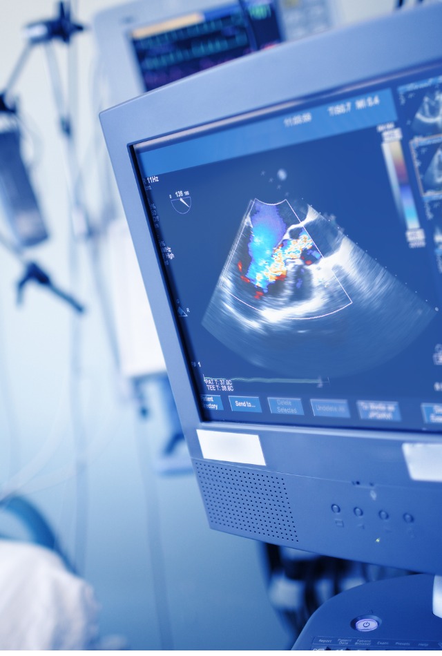
Echocardiography

It is performed for the diagnosis of heart diseases. It is especially used to assess disorders of the heart valves. In this procedure, images of heart valves and muscles are created via ultrasound waves.

Special features of echocardiography

The patient has to lie down bare-chested on the examination table. The gel is put on the chest so that the transducer or probe can move smoothly over the chest. A transducer is a device that is attached to a flexible cable, is actually a modified microphone that directs ultrasound waves into the person’s chest.
The procedure takes approximately 15-30 minutes. It can be done in an outpatient imaging center or at a hospital’s cardiology department.

echocardiography

Different types of echocardiography tests:

Doppler echocardiography – It helps in the evaluation of heart valves’ movement. Also helps to detect the exact obstruction site in blood vessels.

Exercise echocardiography/Stress echo – It is prescribed for the detection of heart problems during exercise, and which are not evident when the body is resting and needs less blood supply. Stress echo is done when heart muscles are strained to provide more blood during exercise.

Trans-esophageal echocardiography (TEE) – It is prescribed in rare cases when regular echocardiography does not provide a clear picture, due to obesity, lung disease, or barrel chest.

Advantages and uses of echocardiography

  • A non-invasive procedure to evaluate heart valves and chambers to diagnose
  • Heart murmurs
  • Endocarditis
  • Improper functioning of the heart/heart muscles leading to heart attack
  • Pericarditis
  • Congenital heart defects/diseases
  • Echocardiography is a safe procedure with little risk or side effects.