
Cardiac Treatment


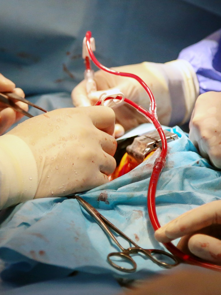
Heart valve replacement
Why is it needed?
It is advised for leaky or narrow heart valve caused by
- Medication
- Calcium deposits
- Infection
- Birth defects
Facts and figures
- Aortic valve was replaced with caged-ball artificial valve done by Hufnagel in year 1952
- Aortic stenosis is the 3rd most common heart disease, usually congenital, occurring in 4 out of 1000 births
- AHA data claims 99,000 heart valve surgery is done per year in the US
Risk and Complications
- Infection
- Bleeding
- Heart attack/stroke
- Breathing problems
- Confusion/ lack of clarity
- Kidney problems
- Arrhythmia
- Reaction to anesthesia or medicine
Advantages
- High success rate
- Successful surgery relieves the patient from weakening symptoms, prevents heart risk failure
- 80% of patients who survive first-year post-surgery can return to normal activity

Disadvantages
- Blood thinners have to be taken by patients using metal valves
- Risk of a malfunctioning or developing leakage, requiring another surgery
- 5% mortality risk
- The long recovery period, especially for older people
Symptoms
- Breathlessness on making slight movements
- Palpitation
- PND – Paroxysmal Nocturnal Dysponea i.e. sudden difficulty in breathing in early morning or at night, awaking the patient
- Black outs – symptom caused by the narrowing of the aortic valves
- Angina
- High temperature
Preoperative Preparation
- Avoid antibiotics and dental procedures pre-surgery
- Full body examination
- Blood and urinalysis
- X-Rays, ECG, stress tests, and cardiac catheterization
- Consult with doctor/surgeon to know which medicines to take or leave out
- Quitting smoking and informing surgeon about alcohol and smoking habits
- The surgeon should be told about a sore throat, cold, fever development before surgery
- Arrange for caregivers post-surgery
- Home should be prepared as advised by the doctor
- No eating or drinking after midnight pre-surgery
- Medical tourists should plan for a long trip and flexible ticket should be booked
Post operative care
- Patient usually stays for 7-10 days in hospital post-surgery, including ICU stay. The patient is constantly monitored.
- Drainage tube is usually removed 1-3 days post surgery
- Blood clot prevention medicines are given
- Patient will feel uncomfortable in the chest while moving about for approx 2 months
- After stabilization, cardiac rehabilitation is started

Dos, Don’ts and Precautions
- Keep the incision hole dry and clean
- Walk daily
- Random blood tests required for patients taking blood thinners
- Change to a healthy lifestyle
- Quit smoking
- Maintain a healthy weight
- Take medicines meticulously
- Take part in a cardiac rehabilitation program
- Regularly visit physician or surgeon
Recovery time is long, especially for the elderly. Health before surgery also has a hand in recovery time. Usually, patients recover fully in 6-8 weeks after surgery.
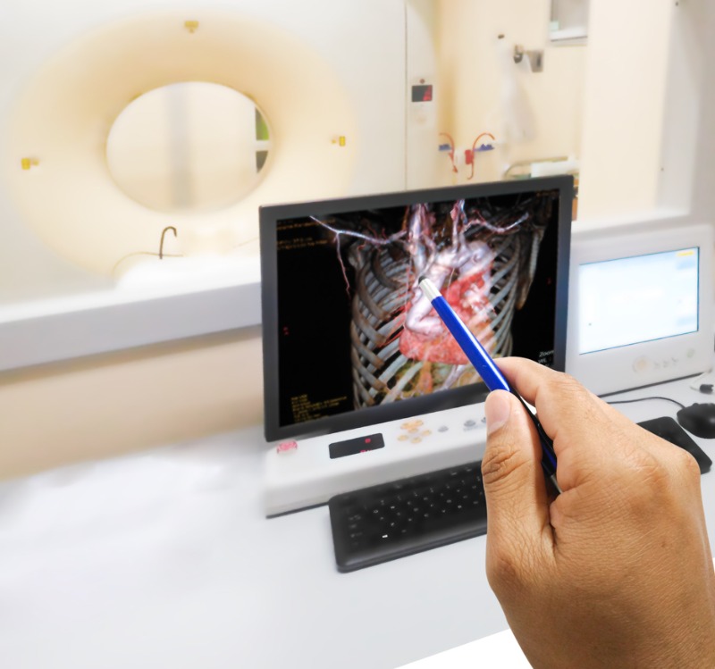
Coronary Angiography
Uses of coronary angiography
- To detect blockage in coronary arteries, and diagnose specific heart diseases such as aortic stenosis, atypical chest pain, unstable angina, unexplained heart failure
- To open blockage if any, using PCI or Percutaneous Coronary Intervention
- To assist physicians in diagnosis as well as to recommend treatment for the relevant coronary diseases
Medical Tourist guide
- Patients can return home 24 hours after the procedure, though hospital stay for 48 hours is advisable. However, if there is any blockage, you can plan for PCI procedure which can happen during the same procedure.
- Coronary angiography is used to find blockages in coronary arteries. Angiography reveals the severity of the coronary disease if any. It helps to select the optimal treatment, which could be coronary stenting, angioplasty, atherectomy, coronary bypass surgery.
Risk and Complications
It is a relatively safe procedure. Some risks associated are:
- Irregular heartbeat
- Low blood pressure
- Arterial damage
- Compression of the heart
- Hemorrhage
- Allergic reaction to the dye
- Infection
- Heart attack\ Stroke
- Kidney damage
Pre Procedure Preparation
- Patient has to be admitted one night before the procedure
- Patient cannot drink or eat anything, 8 hours before procedure
- Inform the doctor about drug allergy/allergies
During the Procedure
- Patient has to wear hospital gown, and will be given mild sedative for comfort and relaxation. Electrodes will be attached to body for heart monitoring purposes
- A catheter will be inserted in the upper thigh or groin, guided by a fluoroscope. The catheter will be advanced up to the opening of the coronary arteries
- Dye is injected via the catheter to coronary artery and several X-Ray images are produced, which are known ad angiograms. The entire process takes approximately 30-60 minutes
- Patient may be awake during the angiography, and feel some pressure when catheter is inserted
- After the procedure is complete, catheter is removed, and insertion hole is sealed or compressed to prevent bleeding
- Patient is transferred to the recovery room, then monitored for about 2-8 hours. The patient has to lie flat on the back in bed for a few hours to prevent bleeding
Coronary Artery Diseases
Quality and lost treatment cost are the prime drivers for foreign medical tourists to India. Heart disease specializing clinics in India operate in a strong and resourceful network making arrangement for following cardiovascular remedies:
Coronary Artery Bypass Graft
This surgical procedure enables sufficient blood transmission to heart carrying oxygen and nutrients. A vein from leg or inner chest wall artery is selected for getting the bypass graft.
Double Valve Replacement
This procedure involves replacement of both the mitral and aortic valve by general anesthesia. Normal patients with acquired heart valve disease or with congenital valve defect are recommended for double valve replacement.
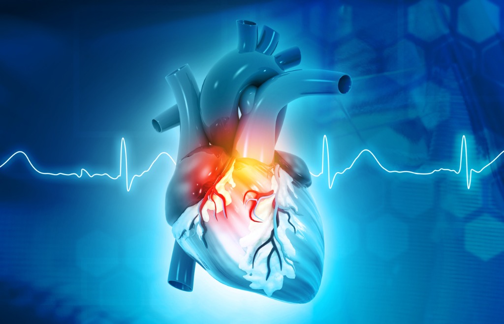
Cardiology
Cardiovascular disorders have taken a menacing form in recent years. It is a prime killer across the world and doesn’t spare anyone on grounds of age. Faulty lifestyle is the chief reason of heart diseases together with genetic precedence and stress. Cardiology Treatment in India handles medical diagnosis and cure of heart-related disorders like:
- Heart failure
- Congenital heart defects
- Electro – physiology and valvular heart ailments
Angiography (including Non – ionic contrast)
The procedure enables an insightful glimpse into the heart chambers, veins, arteries and blood vessels in a non ionic contrast medium.
Angioplasty (including Non – ionic contrast)
A surgery to remove obstructions in coronary artery formed by plaque. The surgery may even be for increasing inner diameter of narrowed blood vessels. For proper visualization of procedure non ionic contrast medium is essential. Two methods instituted are:
- Balloon angioplasty- the heart block removed under the inflated balloon’s impact.
- Balloon stenting – block is removed by placing a balloon stent.
Balloon Aortic Valvuloplasty
- This procedure is done to widen the blocked or narrowed heart valves. A thin flexible tube with a balloon attached is inserted through the arm or groin. As the wire reaches the site, the balloon opens up widening the valve.
Pediatric Cardiac Surgery – Conduit Repair
This stenting procedure covers heart membrane and needed by pediatric patients with congenital heart disorders. A surgery known as atrial switch operation is done to place arteries at the correct position. For sealing the surgical incision, a couple of patches are applied to remedy antrioventricular canal disorder.
Pacemaker Implant Double Chamber (only surgery)
- A small electrical device is set inside the chest to regulate erratic rhythm of the heart. A double chamber pacemaker implant has two links. One connects with the right atrium (upper heart chamber) and the other connects the ventricle (right bottom chamber). The electric pulses in pacemaker sets heart beat to normal rhythm.
Electrophysiology study and Radio frequency catheter ablation
- In the former procedure, electrodes are inserted inside the heart to study defects in heart rhythm and conduction. The later procedure involves sending radio frequency energy to heart. The energy develops heat that melts away the abnormal focus.
CABG (Heart Bypass)
Why did it need?
CABG is prescribed for conditions such as
- The disease of the coronary artery with blockages in 2-3 major coronary arteries
- Chest pain or severe angina is caused by mild exertion
- The function of the left ventricle is poor
- The patient does not respond to any conservative treatment
Facts and figures
- Patients of heart failure have been described in Ebers Papyrus, the oldest, a well-preserved medical document written in approx 1600 BC
- Coronary artery atherosclerosis is the major cause of coronary artery disease. It is the main cause of death in the US
- First CABG was done by surgeons guided by Dr. Robert Goetz
- According to data from AHA, 448, 000 CABG procedures were done in 2006 in the US
- Though the risk is equivalent for all races and genders, the risk increases at age 40 and more
- The mortality rate is more among women, as they develop heart disease 10 years later/after men do
Risk and Complications
- Damage possible to adjacent veins and arteries
- Closing or blockage of graft
- Low or high blood pressure
- Arrhythmia – Abnormal heart rates
- Blood clots may increase heart attack risk
- Mood swings or depression
- Kidney failure
- Anesthesia reaction
- Pulmonary embolism
- Bleeding
- Infection
Advantages
- Marked improvement in ischemia and chest pain
- Surgery helps patients to carry on normal life, apart from improving quality of life
- Prolongs patient life and reduces heart attack risk
- High success rate – 90% of patients are satisfied with CABG
Disadvantages
- CABG cannot prevent coronary artery blockage from recurring
- The overall mortality rate is 4-5%
- Increased lung complication and chest infection risk
- Confusion, short term memory loss is possible, but these subside after about 6 post-surgery
Preoperative Preparation
- Surgery is ideally performed when the patient is stable after a heart attack
- Avoid antibiotics and dental treatment before CABG
- Complete physical examination
- Urine and blood analysis
- Diagnostic tests like stress tests, ECG, X-Rays and cardiac catheterization
- The patient has to consult a doctor or surgeon one week prior to CABG, to obtain list of medicines to be stopped or taken
- Inform surgeon regarding alcohol or smoking habits and quit smoking
- Notify the surgeon if suffering from sore throat, fever or cold before surgery
- Arrange for caregivers after surgery
- Prepare the home as required post-surgery
- The patient should not drink/eat anything prior to surgery
Post operative care
- Patients usually have to stay in the hospital for about 10 days post-surgery, including ICU stay. His/her vital statistics are monitored continuously
- The drainage tube is removed between 1-3 days post-surgery
- The patient will feel discomfort in the chest during any activity for approx 2 months
- Rehabilitation starts after the patient is stable
Symptoms
- Shortness of breath
- Chest pain
- Tiredness, weakness or reduced exertion capacity
- Palpitations, dizziness
- Weight gain, swelling in the leg

Dos, Don’ts and Precautions
- Do keep incision hole dry and clean
- If saphenous vein is used for the graft, the patient should not cross or elevate legs
- Walk daily
- Follow prescribed lifestyle change for maximum benefit
- Quit smoking
- Maintain a healthy weight
- Participate in rehabilitation programs (post-cardiac)
- Do be meticulous in taking medicines
- Do be regular in following up visits to physician and surgeon
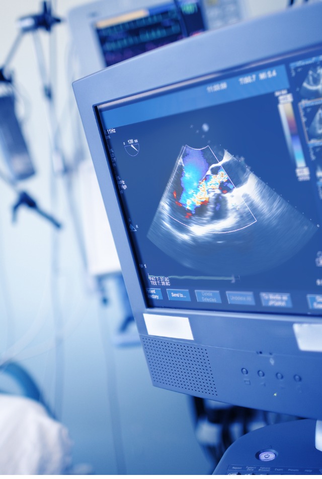
Echocardiography
It is performed for the diagnosis of heart diseases. It is especially used to assess disorders of the heart valves. In this procedure, images of heart valves and muscles are created via ultrasound waves.
Special features of echocardiography
The patient has to lie down bare-chested on the examination table. The gel is put on the chest so that the transducer or probe can move smoothly over the chest. A transducer is a device that is attached to a flexible cable, is actually a modified microphone that directs ultrasound waves into the person’s chest.
The procedure takes approximately 15-30 minutes. It can be done in an outpatient imaging center or at a hospital’s cardiology department.
Different types of echocardiography tests:
Doppler echocardiography – It helps in the evaluation of heart valves’ movement. Also helps to detect the exact obstruction site in blood vessels.
Exercise echocardiography/Stress echo – It is prescribed for the detection of heart problems during exercise, and which are not evident when the body is resting and needs less blood supply. Stress echo is done when heart muscles are strained to provide more blood during exercise.
Trans-esophageal echocardiography (TEE) – It is prescribed in rare cases when regular echocardiography does not provide a clear picture, due to obesity, lung disease, or barrel chest.
Advantages and uses of echocardiography
- A non-invasive procedure to evaluate heart valves and chambers to diagnose
- Heart murmurs
- Endocarditis
- Improper functioning of the heart/heart muscles leading to heart attack
- Pericarditis
- Congenital heart defects/diseases
- Echocardiography is a safe procedure with little risk or side effects.